Other Available Foot and ankle Services
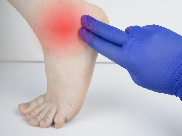
Diabetic Peripheral Neuropathy
What is a Diabetic Peripheral Neuropathy?
Diabetic neuropathy is nerve damage caused by diabetes. When it affects the arms, hands, legs and feet, it is known as diabetic peripheral neuropathy. Diabetic peripheral neuropathy is different from peripheral arterial disease (poor circulation), which affects the blood vessels rather than the nerves.
Three different groups of nerves can be affected by diabetic neuropathy:
- Sensory nerves, which enable people to feel pain, temperature and other sensations
- Motor nerves, which control the muscles and give them their strength and tone
- Autonomic nerves, which allow the body to perform certain involuntary functions, such as sweating
Diabetic Peripheral Neuropathy Causes
The nerve damage that characterizes diabetic peripheral neuropathy is more common in patients with poorly managed diabetes. However, even patients living with diabetes who have excellent blood sugar (glucose) control can develop diabetic neuropathy. There are several theories as to why this occurs, including the possibilities that high blood glucose or constricted blood vessels produce damage to the nerves.
As diabetic peripheral neuropathy progresses, various nerves are affected. These damaged nerves can cause problems that encourage development of ulcers. For example:
- Deformities (such as bunions or hammertoes) resulting from motor neuropathy may cause shoes to rub against toes, creating a sore. The numbness caused by sensory neuropathy can make the patient unaware that this is happening.
- Because of numbness, a patient may not realize that s/he has stepped on a small object and cut the skin.
Cracked skin caused by autonomic neuropathy, combined with sensory neuropathy’s numbness and problems associated with motor neuropathy, can lead to developing a sore.
Motor Neuropathy (Deformity) + Ill-Fitting Shoes + Sensory Neuropathy (numbness) = Ulcers (sores)
Diabetic Peripheral Neuropathy Symptoms
Depending on the type(s) of nerves involved, one or more symptoms may be present in diabetic peripheral neuropathy.
For sensory neuropathy:
- Numbness or tingling in the feet
- Pain or discomfort in the feet or legs, including prickly, sharp pain or burning feet
For motor neuropathy:
- Muscle weakness and loss of muscle tone in the feet and lower legs
- Loss of balance
- Changes in foot shape that can lead to areas of increased pressure
For autonomic neuropathy:
- Dry feet
- Cracked skin
Diabetic Peripheral Neuropathy Diagnosis
To diagnose diabetic peripheral neuropathy, the foot and ankle surgeon will obtain the patient’s history of symptoms and will perform simple in-office tests on the feet and legs. This evaluation may include assessment of the patient’s reflexes, ability to feel light touch and ability to feel vibration. In some cases, additional neurologic tests may be ordered.
INGROWN TOENAILS
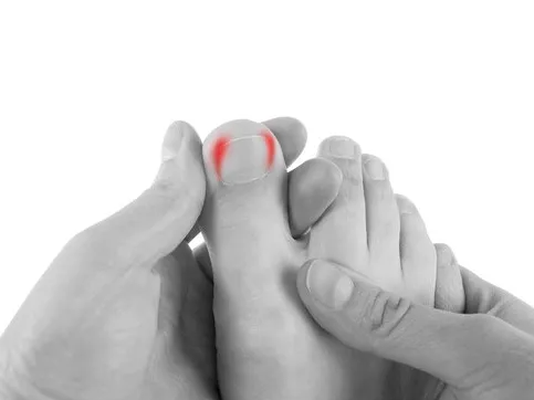
What is an Ingrown Toenail?
When a toenail is ingrown, the nail is curved downward and grows into the skin, usually at the nail borders (the sides of the nail). This “digging in” of the nail irritates the skin, often creating pain, redness, swelling, and warmth in the toe.
If an ingrown nail causes a break in the skin, bacteria may enter and cause an infection in the area, which is often marked by drainage and a foul odor. However, even if your toe isn’t painful, red, swollen, or warm, a nail that curves downward into the skin can progress to an infection.
Ingrown Nail Causes
Ingrown toenails can develop for various reasons. In many people, the tendency to have this common disorder is inherited. In other cases, an ingrown toenail is the result of trauma, such as stubbing your toe, having an object fall on your toe, or engaging in activities that involve repeated pressure on the toes, such as kicking or running.
The most common cause of ingrown toenails is improper trimming. Cutting your nails too short encourages the skin next to the nail to fold over the nail. Another cause of ingrown toenails is wearing shoes that are tight or short.
Certain nail conditions are often associated with ingrown toenails. For example, if you have had a toenail fungal infection or if you have lost a nail through trauma, you are at greater risk for developing an ingrown toenail.
Ingrown Toenail Treatment
Sometimes initial treatment for ingrown toenails can be safely performed at home. However, home treatment is strongly discouraged if you suspect you have an infection, or if you have a medical condition that puts your feet at high risk—for example, diabetes, nerve damage in the foot, or poor circulation.
Ingrown Toenail Prevention
Many cases of ingrown toenails may be prevented by following these two important tips:
- Trim your nails properly. Cut your toenails in a fairly straight line, and don’t cut them too short. You should be able to get your fingernail under the sides and end of the nail.
- Avoid poorly-fitting shoes. Don’t wear shoes that are short or tight in the toe box. Also avoid shoes that are loose, because they too cause pressure on the toes, especially when you run or walk briskly.
HEEL PAIN & PLANTAR FASCIITIS
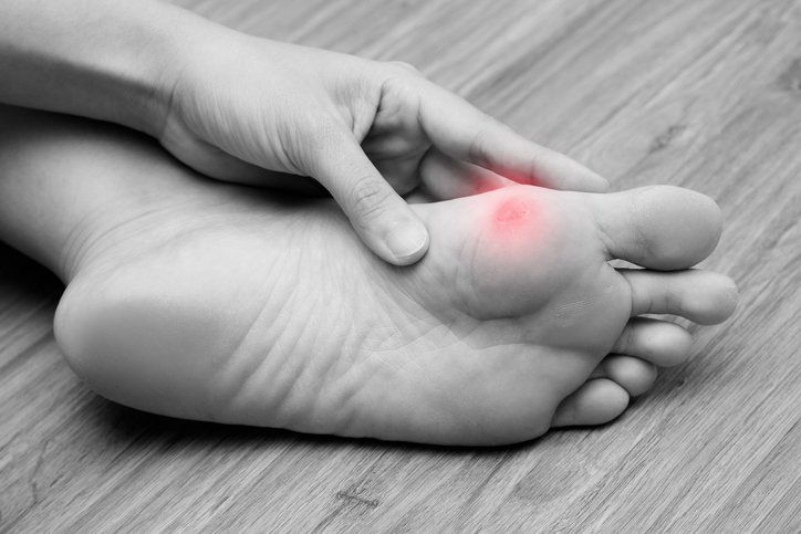
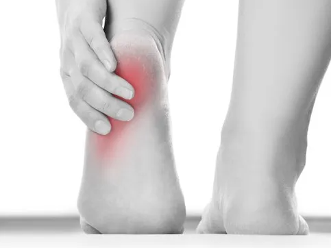
What is a Plantar Fasciitis?
Plantar fasciitis is an inflammation of the band of tissue (the plantar fascia) that extends from the heel to the toes. In this condition, the fascia first becomes irritated and then inflamed, resulting in heel pain.
Plantar Fasciitis Causes
The most common cause of plantar fasciitis relates to faulty structure of the foot. For example, people who have problems with their arches, either overly flat feet or high-arched feet, are more prone to developing plantar fasciitis.
Wearing nonsupportive footwear on hard, flat surfaces puts abnormal strain on the plantar fascia and can also lead to plantar fasciitis. This is particularly evident when one’s job requires long hours on the feet. Obesity and overuse may also contribute to plantar fasciitis.
Plantar Fasciitis Symptoms
The symptoms of plantar fasciitis are:
- Pain on the bottom of the heel
- Pain in the arch of the foot
- Pain that is usually worse upon arising
- Pain that increases over a period of months
- Swelling on the bottom of the heel
People with plantar fasciitis often describe the pain as worse when they get up in the morning or after they have been sitting for long periods of time. After a few minutes of walking, the pain decreases because walking stretches the fascia. For some people, the pain subsides but returns after spending long periods of time on their feet.
Plantar Fasciitis Diagnosis
To arrive at a diagnosis, the foot and ankle surgeon will obtain your medical history and examine your foot. Throughout this process, the surgeon rules out all possible causes for your heel pain other than plantar fasciitis.
In addition, diagnostic imaging studies, such as x-rays or other imaging modalities, may be used to distinguish the different types of heel pain. Sometimes heel spurs are found in patients with plantar fasciitis, but these are rarely a source of pain. When they are present, the condition may be diagnosed as plantar fasciitis/heel spur syndrome.
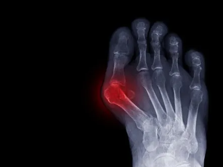
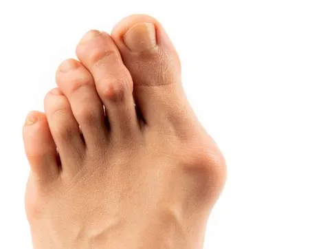
BUNIONS
What is a Bunion?
A bunion (also referred to as hallux valgus or hallux abducto valgus) is often described as a bump on the side of the big toe. But a bunion is more than that. The visible bump actually reflects changes in the bony framework of the front part of the foot. The big toe leans toward the second toe, rather than pointing straight ahead. This throws the bones out of alignment – producing the bunion’s “bump.”
Bunions are a progressive disorder. They begin with a leaning of the big toe, gradually changing the angle of the bones over the years and slowly producing the characteristic bump, which becomes increasingly prominent. Symptoms usually appear at later stages, although some people never have symptoms.
Bunion Causes
Bunions are most often caused by an inherited faulty mechanical structure of the foot. It is not the bunion itself that is inherited, but certain foot types that make a person prone to developing a bunion. Although wearing shoes that crowd the toes won’t actually cause bunions, it sometimes makes the deformity get progressively worse. Symptoms may therefore appear sooner.
Bunion Symptoms
Symptoms, which occur at the site of the bunion, may include:
- Pain or soreness
- Inflammation and redness
- A burning sensation
- Possible numbness
Symptoms occur most often when wearing shoes that crowd the toes, such as shoes with a tight toe box or high heels. This may explain why women are more likely to have symptoms than men. In addition, spending long periods of time on your feet can aggravate the symptoms of bunions.
Bunion Diagnosis
Bunions are readily apparent – the prominence is visible at the base of the big toe or side of the foot. However, to fully evaluate the condition, the foot and ankle surgeon may take x-rays to determine the degree of the deformity and assess the changes that have occurred.
HAMMERTOES
What is a Hammertoe?
Hammertoe is a contracture (bending) deformity of one or both joints of the second, third, fourth or fifth (little) toes. This abnormal bending can put pressure on the toe when wearing shoes, causing problems to develop.
Normal toes unaffected by Hammertoes usually start out as mild deformities and get progressively worse over time. In the earlier stages, hammertoes are flexible and the symptoms can often be managed with noninvasive measures. But if left untreated, hammertoes can become more rigid and will not respond to nonsurgical treatment.
Because of the progressive nature of hammertoes, they should receive early attention. Hammertoes never get better without some kind of intervention.
Hammertoe Causes
The most common cause of hammertoe is a muscle/tendon imbalance. This imbalance, which leads to a bending of the toe, results from mechanical (structural) or neurological changes in the foot that occur over time in some people.
HammertoesHammertoes may be aggravated by shoes that do not fit properly. A hammertoe may result if a toe is too long and is forced into a cramped position when a tight shoe is worn. Occasionally, hammertoe is the result of an earlier trauma to the toe. In some people, hammertoes are inherited.
Hammertoe Symptoms
Common symptoms of hammertoes include:
- Pain or irritation of the affected toe when wearing shoes.
- Corns and calluses (a buildup of skin) on the toe, between two toes or on the ball of the foot. Corns are caused by constant friction against the shoe. They may be soft or hard, depending on their location.
- Inflammation, redness or a burning sensation
- Contracture of the toe
- In more severe cases of hammertoe, open sores may form.
Hammertoe Diagnosis
Although hammertoes are readily apparent, to arrive at a diagnosis, the foot and ankle surgeon will obtain a thorough history of your symptoms and examine your foot. During the physical examination, the doctor may attempt to reproduce your symptoms by manipulating your foot and will study the contractures of the toes. In addition, the foot and ankle surgeon may take x-rays to determine the degree of the deformities and assess any changes that may have occurred.
Hammertoes are progressive—they do not go away by themselves and usually they will get worse over time. However, not all cases are alike—some hammertoes progress more rapidly than others. Once your foot and ankle surgeon has evaluated your hammertoes, a treatment plan can be developed that is suited to your needs.
ANKLE SPRAINS
What is an Ankle Sprain?
An ankle sprain is an injury to one or more ligaments in the ankle, usually on the outside of the ankle. Ligaments are bands of tissue—like rubber bands—that connect one bone to another and bind the joints together. In the ankle joint, ligaments provide stability by limiting side-to-side movement.
Some ankle sprains are much worse than others. The severity of an ankle sprain depends on whether the ligament is stretched, partially torn or completely torn, as well as on the number of ligaments involved. Ankle sprains are not the same as strains, which affect muscles rather than ligaments.
Ankle Sprain Causes
Sprained ankles often result from a fall, a sudden twist or a blow that forces the ankle joint out of its normal position. Ankle sprains commonly occur while participating in sports, wearing inappropriate shoes or walking or running on an uneven surface.
Sometimes ankle sprains occur because a person is born with weak ankles. Previous ankle or foot injuries can also weaken the ankle and lead to sprains.
Ankle Sprain Symptoms
The symptoms of ankle sprains may include:
- Pain or soreness
- Swelling
- Bruising
- Difficulty walking
- Stiffness in the joint
Ankle fractures are common injuries most often caused by the ankle rolling inward or outward. Many people mistake an ankle fracture for an ankle sprain, but they are quite different and therefore require an accurate and early diagnosis. They sometimes occur simultaneously.
Ankle Sprain Diagnosis
In evaluating your injury, the foot and ankle surgeon will obtain a thorough history of your symptoms and examine your foot. X-rays or other advanced imaging studies may be ordered to help determine the severity of the injury.
Why Prompt Medical Attention is Needed
There are four key reasons why an ankle sprain should be promptly evaluated and treated by a foot and ankle surgeon:
- An untreated ankle sprain may lead to chronic ankle instability, a condition marked by persistent discomfort and a giving way of the ankle. Weakness in the leg may also develop.
- A more severe ankle injury may have occurred along with the sprain. This might include a serious bone fracture that, if left untreated, could lead to troubling complications.
- An ankle sprain may be accompanied by a foot injury that causes discomfort but has gone unnoticed thus far.
- Rehabilitation of a sprained ankle needs to begin right away. If rehabilitation is delayed, the injury may be less likely to heal properly.

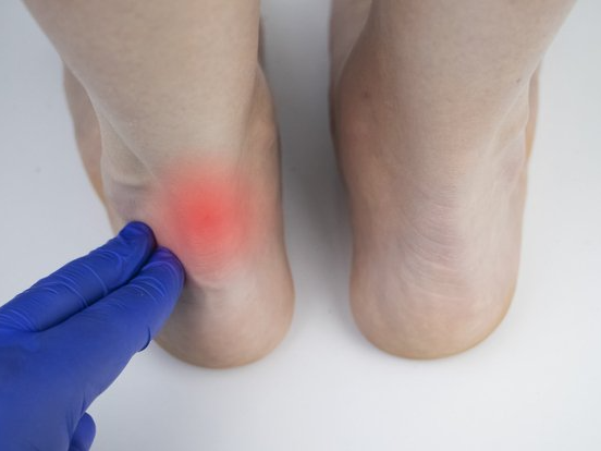
ACHILLES TENDON DISORDERS
What is the Achilles Tendon?
The Achilles tendon is a band of tissue that connects a muscle to a bone. It runs down the back of the lower leg and connects the calf muscle to the heel bone. Also called the heel cord, the Achilles tendon facilitates walking by helping to raise the heel off the ground.
Two common disorders that occur in the heel cord are Achilles tendonitis and Achilles tendonosis.
Achilles tendonitis is an inflammation of the Achilles tendon. This inflammation is typically short-lived. Over time, if not resolved, the condition may progress to a degeneration of the tendon (Achilles tendonosis), in which the tendon loses its organized structure and is likely to develop microscopic tears. Sometimes the degeneration involves the site where the Achilles tendon attaches to the heel bone. In rare cases, chronic degeneration with or without pain may result in rupture of the tendon.
Achilles Tendon Disorder Causes
As "overuse" disorders, Achilles tendonitis and tendonosis are usually caused by a sudden increase of a repetitive activity involving the Achilles tendon. Such activity puts too much stress on the tendon too quickly, leading to micro-injury of the tendon fibers. Due to this ongoing stress on the tendon, the body is unable to repair the injured tissue. The structure of the tendon is then altered, resulting in continued pain.Diagram of side of foot showing zone of tendonitis or tendonosis
Athletes are at high risk for developing disorders of the Achilles tendon. Achilles tendonitis and tendonosis are also common in individuals whose work puts stress on their ankles and feet, such as laborers, as well as in “weekend warriors”—those who are less conditioned and participate in athletics only on weekends or infrequently.
In addition, people with excessive pronation (flattening of the arch) have a tendency to develop Achilles tendonitis and tendonosis due to the greater demands placed on the tendon when walking. If these individuals wear shoes without adequate stability, their overpronation could further aggravate the Achilles tendon.
Achilles Tendonitis & Tendonosis Symptoms
The symptoms associated with Achilles tendonitis and tendonosis include:
- Pain—aching, stiffness, soreness or tenderness—within the tendon. This may occur anywhere along the tendon’s path, beginning with the tendon’s attachment directly above the heel upward to the region just below the calf muscle. Pain often appears upon arising in the morning or after periods of rest, then improves somewhat with motion but later worsens with increased activity.
- Tenderness, or sometimes intense pain, when the sides of the tendon are squeezed. There is less tenderness, however, when pressing directly on the back of the tendon.
- When the disorder progresses to degeneration, the tendon may become enlarged and may develop nodules in the area where the tissue is damaged.
Achilles Tendonitis or Tendonosis Diagnosis
In diagnosing Achilles tendonitis or tendonosis, the surgeon will examine the patient’s foot and ankle and evaluate the range of motion and condition of the tendon. The extent of the condition can be further assessed with x-rays or other imaging modalities.
PERONEAL TENDON INJURIES
What are the Peroneal Tendons?
A tendon is a band of tissue that connects a muscle to a bone. The two peroneal tendons in the foot run side by side behind the outer ankle bone. One peroneal tendon attaches to the outer part of the midfoot, while the other tendon runs under the foot and attaches near the inside of the arch. The main function of the peroneal tendons is to stabilize the foot and ankle and protect them from sprains.
Causes of Peroneal Tendon Injuries
Peroneal tendon injuries may be acute (occurring suddenly) or chronic (developing over a period of time). They most commonly occur in individuals who participate in sports that involve repetitive ankle motion. In addition, people with higher arches are at risk for developing peroneal tendon injuries. Basic types of peroneal tendon injuries are tendonitis, tears and subluxation.
Tendonitis is an inflammation of one or both tendons. The inflammation is caused by activities involving repetitive use of the tendon, overuse of the tendon or trauma (such as an ankle sprain).
Acute tears are caused by repetitive activity or trauma. As time goes on, these tears may lead to a change in the shape of the foot in which the arch may become higher.
Degenerative tears (tendonosis) are usually due to overuse and occur over long periods of time, often years. In degenerative tears, the tendon is like taffy that has been overstretched until it becomes thin and eventually frays. Having high arches also puts you at risk for developing a degenerative tear.
Subluxation means one or both tendons have slipped out of their normal position. In some cases, subluxation is due to a condition in which a person is born with a variation in the shape of the bone or muscle. In other cases, subluxation occurs following trauma, such as an ankle sprain. Damage or injury to the tissues that stabilize the tendons (retinaculum) can lead to chronic tendon subluxation.
Early treatment of a subluxation is critical since a tendon that continues to sublux (move out of position) is more likely to tear or rupture. Therefore, if you feel the characteristic snapping, see a foot and ankle surgeon immediately
Symptoms of Peroneal Tendon Injuries
Tendonitis:
- Pain
- Swelling
- Warm to the touch
Acute tears:
- Pain
- Swelling
- Weakness or instability of the foot and ankle
Degenerative tears (tendonosis):
- Sporadic pain (occurring from time to time) on the outside of the ankle
- Weakness or instability in the ankle
- An increase in the height of the arch
Subluxation:
- A snapping feeling of the tendon around the ankle bone
- Sporadic pain behind the outside ankle bone
- Ankle instability or weakness
Peroneal Tendon Injury Diagnosis
Because peroneal tendon injuries are sometimes misdiagnosed and may worsen without proper treatment, prompt evaluation by a foot and ankle surgeon is advised. To diagnose a peroneal tendon injury, the surgeon will examine the foot and look for pain, instability, swelling, warmth and weakness on the outer side of the ankle. In addition, an x-ray or other advanced imaging studies may be needed to fully evaluate the injury. The foot and ankle surgeon will also look for signs of an ankle sprain and other related injuries that sometimes accompany a peroneal tendon injury. Proper diagnosis is important because prolonged discomfort after a simple sprain may be a sign of additional problems.